What types of knee preservation techniques are available?
Therefore, a comprehensive and individualized treatment plan that takes into account a person’s age, activity level, anticipated and degree of joint dysfunction is an important consideration for joint preservation. This is a matter. Multidisciplinary treatment options are often part of a comprehensive plan and include physical activity modifications, physical therapy, medications, injections, and conservative surgery.
What are the non-surgical approaches to preserving the knee joint?
Physical Therapy: Lack of physical activity often leads to joint damage, and physical therapy and exercise are part of the defense against such damage. It’s on the front line. It can be difficult to move during an acute pain episode, and inappropriate exercise choices can cause even more damage. Therefore, an individualized exercise program under the supervision of a qualified physical therapist or orthopedic surgeon can help restore function and reduce pain.
Exercise: A customized exercise plan not only improves a person’s overall health, but also helps strengthen and increase mobility of the muscles, ligaments, and tissues in the injured area.
Analgesic modalities such as IFT, ultrasound: Local application of analgesic modalities such as ice, massage, deep heat, and sometimes nerve stimulation and ultrasound have proven helpful in some cases. .
Lifestyle changes: A sedentary and stressful lifestyle can have a negative impact on a person’s overall health and bone health. Quitting harmful lifestyle habits can help preserve your knee joints. Some approaches are:
Weight Loss: As mentioned earlier, the knee joint takes the brunt of excess body weight, so it has to work harder. Weight management strategies under the supervision of a qualified physician can make a big difference in removing harmful forces from the knee joint.
Yoga, Meditation, and Dietary Changes: Relaxation through lifestyle management techniques such as yoga, meditation, and dietary management strategies under the supervision of a qualified nutritionist can help reduce knee joint pain.
Simple Painkillers, NSAIDs: Nonsteroidal anti-inflammatory drugs available over the counter can help relieve acute pain and swelling in the knee joint. Taking such drugs for a long time can be harmful, so they should only be taken under the supervision of a doctor or orthopedic surgeon.
Chondroprotectors: Chemicals naturally produced in the body, such as glucosamine and chondroitin, are commercially available as packaged dietary supplements. These may also be prescribed by your doctor. Glucosamine plays a role in repairing articular cartilage, while chondroitin prevents certain other enzymes from destroying articular cartilage.
Relief Orthoses: External aids such as knee braces and knee bandages may be beneficial in some cases by reducing pressure and increasing stability. Additionally, assistive devices such as walkers, canes, and crutches take weight off arthritic knees. This reduces pain when walking or straining, and also reduces the risk of falls.
Injections into the join :
Steroids: If NSAIDs do not produce the desired results, corticosteroids may be prescribed in the form of injections into the joint space to reduce inflammation. Corticosteroid analgesia results from the stimulatory effect of steroids on prostaglandin synthesis. Steroids also reduce the activity of collagenase and other cartilage-destroying enzymes.
Viscous Replenishment with Hyaluronic Acid: Synovial fluid containing hyaluronic acid is the body’s own lubricant in joint cavities. Joint pain can be a symptom of hyaluronic acid deficiency. Topically supplementing this acid in the form of an injection cannot stop the progression of arthritis, but it can relieve pain and stiffness in some cases.
Prolotherapy (Regenerative Injection Therapy or Prolotherapy): Injecting a natural irritant, often a type of sugar called glucose, into the soft tissues of an injured knee joint is called prolotherapy. Stimulants trigger a healing response in the body.
PRP / Adipose Tissue / BMAC: Concentrated platelets from human blood are called platelet-rich plasma (PRP). In addition to having blood-clotting properties, platelets also contain proteins called growth factors that the body uses to heal injuries. PRP may be used as a joint-sparing approach when more conservative options such as physical therapy, bracing, and activity modification are ineffective.
Radiofrequency ablation (or RFA): This technique involves applying electrical current from radiofrequency waves to heat a small area of nerve tissue around the affected knee joint. This weakens pain signals from the affected area. RFA is generally well tolerated and rarely causes complications.
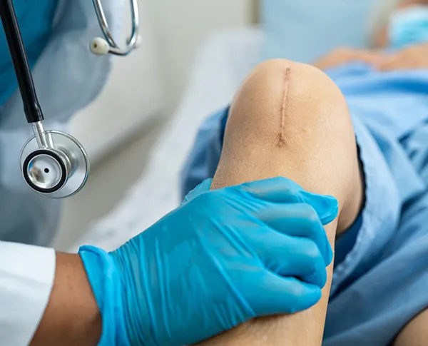
What surgical methods are available to preserve the knee joint ?
Joint preservation can be achieved by minimally invasive surgical techniques in combination with conservative approaches, or when conservative approaches fail. can. Surgical approaches include:
1. Joint realignment procedures: Wear or arthritic areas of the knee joint can be alleviated by surgically correcting leg misalignment using realignment procedures. This reduces pressure on the worn parts of the knee and reduces pain. Realignment can be done in the following ways:
2. Proximal fibular osteotomy: A proximal fibular osteotomy involves cutting and reshaping the fibula (fibula) to relieve pressure on the knee joint. It is a minimally invasive day treatment procedure that includes: This is a preferred alternative for relieving osteoarthritis pain on the medial side of the knee joint. The load is transferred to the lateral (lateral) compartment and the medial (medial) compartment of the knee is relieved. This not only reduces the load on the medial part, but also expands the joint space. Patients can stand and walk the afternoon of surgery, climb stairs the next day, and be able to stand for 1 to 2 hours within 3 to 4 days. The main advantage of this technique is its simplicity and ease of implementation. The procedure can be performed with a short incision without damaging adjacent tissues. This surgery does not include internal implants. All these reasons also result in a shorter recovery period.
3. High Tibial Osteotomy: The knee may bear weight asymmetrically, but usually on the medial or medial portion of the joint (valgus or varus malalignment). As a result, more wear occurs on the medial side of the knee joint than on the outside (outside). In such cases, the legs begin to look bowlegged. In these specific cases, a high tibial osteotomy, a surgical procedure that realigns the knee joint, is required. In this surgery, part of the tibia (shinbone) is cut and reshaped to relieve pressure on the knee joint. The benefits are similar to proximal fibular osteotomy.
4. Microfracture: In the Microfracture Surgical Technique for Articular Cartilage Repair, small fractures are created in the underlying bone. A super clot forms at the fracture site and stimulates the development of new cartilage. Surgery is beneficial because it is quick, less invasive, and has a short recovery time.
Autologous chondrocyte implantation (ACI) is a surgical technique used to treat isolated full-thickness articular cartilage defects in the knee. In this procedure, an orthopedic surgeon removes a small piece of articular cartilage from a patient’s knee. The cartilage is treated with enzymes in the laboratory to isolate cartilage-producing cells, or chondrocytes. These cells are grown in culture and transplanted into the affected area after a few weeks. During implantation, a small patch is sewn over the articular cartilage defect and cells are injected underneath this patch. The cells then grow and form new hyaline cartilage, which resembles natural articular cartilage.
5. Arthroscopic OATS (Osteoarticular Transfer System): A surgical procedure indicated for the treatment of localized cartilage defects. Cartilage is removed from the undamaged area of the joint and surgically placed into the damaged area.
Ligament Reconstruction in Knee Joint Preservation ?
1. Anterior Cruciate Ligament (ACL) Reconstruction: This technique is performed to repair tears in the anterior cruciate ligament, an important stabilizing ligament in the knee. To do this, the surgeon uses tissue around the patella or quadriceps. Thanks to recent advances in arthroscopic technology, ACL surgery can now be performed as a minimally invasive procedure with a low complication rate.
2. PCL Reconstruction: The posterior cruciate ligament (posterior cruciate ligament) is also the main ligament in the knee that connects the femur (thigh bone) to the tibia (shin bone). The PCL limits the posterior movement of the tibia. Surgery is generally considered for people who have dislocated their knee and have multiple ligament tears, including a torn posterior cruciate ligament. During surgery, tissue grafts from another part of the body are used to rebuild torn ligaments.
3. Meniscal Repair: The meniscus is a piece of cartilage that acts as a cushion between the femoral joint and the tibia bone. This cartilage is often damaged by injury or wear and tear. Meniscal tear repair can be done endoscopically or through keyhole surgery.
4. Debridement + Irrigation: Excessive accumulation of inflammatory fluid within the knee joint can cause pain. Visually guided introduction of saline into the knee joint by rinsing or irrigation helps remove this fluid and any loose bodies that may be present within the knee joint. In addition to irrigation, the orthopedic surgeon may also perform surgical debridement and smoothing of the bone surface.
5. Partial Knee Replacement: In this procedure, only a part of the knee is replaced instead of the whole knee if the defect is limited only to a single area of the knee and the rest of the bone is healthy.
Postoperative Rehabilitation After Knee-Sparing Surgery ?
Joint-sparing surgery, when combined with a thorough postoperative rehabilitation program and regular physical therapy, maintains range of motion, achieves muscle reactivation, and reduces postoperative swelling. very effective in reducing.
Cartilage repair procedures typically take at least 6 to 8 weeks to heal and repair. In some cases, additional aids such as crutches may be needed for several weeks. Improving range of motion and increasing muscle strength will begin after two weeks, and he will be able to begin high-intensity physical activity, such as running, after four to six months when the bones have healed sufficiently.
The success of joint-sparing surgery is also highly dependent on the person who develops an individualized treatment plan with a physical therapist after surgery. Maximum positive results can only be achieved if a properly managed rehabilitation program is followed. Otherwise, stiffness, scarring, and muscle atrophy may return.
Actual recovery time will vary from person to person and depends on a variety of factors, including: The person’s underlying health status and preservation techniques used.
Is Knee-sparing surgery safe ? What are the risks ?
In general, knee-sparing surgery is a safe procedure with relatively low risks and a low complication rate. However, as with any surgery, there may be general and individual risks. Common risks include:
- Accidental damage to surrounding structures
- Infection
- Bleeding
- Pain and swelling
- Side effects of anesthesia
How should one choose a facility for knee joint preservation surgery in Nagpur ?
Minimal invasive joint preservation surgery requires modern and innovative techniques of orthopedic surgery and sophisticated equipment. The approach towards joint preservation should be multidisciplinary including minimally invasive surgical, non-surgical and rehabilitative modalities.
The knee joint being a weight-bearing joint is subjected to lifelong pressure; as a result, it is very vulnerable to injuries and wear-tears. One of the main objectives of joint preservation is to relieve the person of pain, restore the natural functioning of the joint and avoid collateral damage to the adjacent tissues. This objective can be achieved by a precise identification of the underlying defect, sophisticated equipment used in the techniques, and surgical expertise which is usually not available in all healthcare settings. Hence the treatment is available only at a few select super-specialty hospitals in India.
The decision to have knee replacement surgery or preservation of the joint is an important one that needs to be seriously taken collectively by the affected person, family, and orthopedic surgeon. Finding the right facility and surgeon is paramount for successful outcomes and also to ensure that the person is comfortable with the decision to have surgery if needed.