
Dr. Sanjay Barik
Knee & Shoulder Surgeon
Dr. Sanjay Barik
Knee & Shoulder Surgeon


Meet Our Doctor
Dr. Sanjay Barik
Orthopedic and Joint Replacement Surgeon
Dr. Barik's Orthocare Clinic
- MBBS
- MS - Orthopaedics
Best Shoulder Arthroscopy In Nagpur
Shoulder arthroscopy:
Shoulder arthroscopy is a minimally invasive surgical procedure that has become a valuable tool in orthopedics for diagnosing and treating various problems related to the shoulder joint. This procedure is often used to treat conditions such as rotator cuff tears, shoulder impingement, labral tears, and other joint abnormalities.
Here is a detailed breakdown of the main aspects of shoulder arthroscopy:
1. Indications:
Rotator cuff tears: Arthroscopy stabilizes and stabilizes groups of muscles and tendons. It is often used to repair rotator cuff tears. For the movement of the shoulder joint.
Shoulder Impingement: This occurs when the rotator cuff tendons become wedged between the bones of the shoulder, causing pain and inflammation.
Labrum tear: The labrum is the rim of cartilage that surrounds the shoulder socket. Tears of the labrum can be treated using arthroscopic techniques.
Joint infections: Arthroscopy helps diagnose and treat joint infections, allowing for targeted interventions.
2. Procedure:
The patient is usually placed under general anesthesia and the surgeon makes a small incision around the shoulder.
An arthroscope, a thin tube with a light source and a camera, is inserted through one of the incisions to visualize the inside of the joint.
Additional small incisions are made to insert special surgical instruments to repair or treat the identified problem.
Surgeons can accurately examine joints, repair damaged tissue, remove debris, and perform other necessary procedures.
3. Advantages of Shoulder Arthroscopy:
Minimally Invasive: The procedure involves small incisions, reducing trauma to surrounding tissues compared to traditional open surgery.
Faster recovery: Patients typically experience less pain and faster recovery after surgery.
Diagnostic Accuracy: Arthroscopy allows direct visualization of joints, allowing for accurate diagnosis and targeted treatment.
Reduced scarring: Smaller incisions minimize scarring and improve cosmetic results.
4. Postoperative Care:
After surgery, patients may receive physical therapy to restore shoulder strength and mobility.
Specific postoperative instructions will be provided, including restrictions on certain activities during the initial recovery period.
Follow-up appointments with your surgeon are essential to monitor your progress and address any concerns.
5. Continuous Advancement:
Shoulder arthroscopy technology is constantly evolving, and so are the instruments and procedures.
New technologies such as 3D imaging and improved arthroscopic instruments are contributing to improved surgical outcomes.
In summary, shoulder arthroscopy has revolutionized the approach to diagnosis and treatment of various diseases of the shoulder joint. Minimally invasive, combined with continued advances in technology, patients can benefit from accurate diagnosis, targeted treatment, and faster recovery. This procedure has significantly improved the overall patient experience when treating shoulder problems in the orthopedic field.
Anatomy:
The shoulder is a complex joint that can perform more movements than any other joint in the human body. It is made up of three bones:
- Bone of the humerus (humerus)
- Scapula (scapula)
- Clavicle (clavicle)
Anatomy of the ball and socket joint of the shoulder:
The shoulder joint is a ball and socket joint. The joint socket where the head of the upper arm bone (humerus) fits into the round socket (glenoid fossa) of the scapula. Articular cartilage covers the surface of the ball and socket and ensures smooth, smooth movement.
Labrum and Stability:
The glenoid fossa is surrounded by the labrum, a fibrocartilage that provides stability and acts as a cushion for the joint.
Shoulder Capsule and Synovium:
The ligaments form a capsule around the joint, and a thin membrane, the synovium, lines the underside of the capsule. The synovium produces synovial fluid that lubricates joints and ensures smooth movement.
Rotator Cuff and Tendons:
Four tendons, collectively called the rotator cuff, surround the shoulder capsule. These tendons help center the arm bone in the fossa, cover the humeral head, and attach it to the scapula.
Bursae for smooth movement:
A sliding sac called the bursa is located between the rotator cuff and the acromion (the top bone of the shoulder). The bursa facilitates the smooth glide of the rotator cuff tendons as you move your arm.
Shoulder Arthroscopy Indications:
Shoulder arthroscopy is recommended when a painful condition cannot be resolved by non-surgical treatments such as rest, physical therapy, and drug therapy. Common problems include rotator cuff tendon injuries, labral tears, and inflammation that affects the soft tissues around the joint.
General Arthroscopic Surgery:
Rotator Cuff Repair: Treatment of tears or injuries to the rotator cuff tendons.
Labrum removal or repair: Treatment of tears or damage to the periglenoid fibrocartilage.
Ligament Repair: Corrects problems in the ligaments that form the joint capsule.
Tissue Removal or Cartilage Repair: Removal of inflamed tissue or repair of damaged articular cartilage.
Treatment of chronic shoulder instability: Repair of recurrent shoulder dislocations.
Less Common Arthroscopy:
In addition to common procedures, arthroscopy can also be used for less common problems such as nerve release, fracture repair, and cyst removal. However, certain surgeries, such as shoulder replacements, may still require traditional open surgery.
In summary, shoulder arthroscopy is a minimally invasive surgery that treats a variety of problems in the shoulder joint using small incisions and advanced techniques to diagnose, treat, and repair damaged tissue. This can be a valuable option when non-surgical treatments prove ineffective in treating painful shoulder conditions.
Planning Your Surgery
Reviews and Tests
Your orthopedic surgeon may request that you see your primary care physician to ensure that there are no medical issues that need to be addressed before surgery. Blood tests, an electrocardiogram, or a chest x-ray may be required to ensure the surgery is safe.
If you have certain health risks, you may need a more detailed evaluation before surgery.
Precautions for hospitalization
If you are generally healthy, arthroscopy will likely be performed on an outpatient basis. This means you don’t need to stay in the hospital overnight.
Be sure to tell your orthopedic surgeon about any medications or supplements you are taking. You may need to stop taking some of these medications before surgery.
The hospital or surgery center will contact you in advance for further details about your surgery. Before your surgery, be sure to follow the instructions regarding your arrival time and especially when to stop eating and drinking.
Anesthesia
Before your surgery, our anesthesia staff will discuss your anesthesia options with you.
Shoulder arthroscopy can be performed using a local nerve block to numb the shoulder and arm. This numbing drug is injected high into the neck or shoulder. This is where the nerves that control sensation in your shoulders and arms are located. Nerve blocks are not only used as an anesthetic during surgery, but they can also help reduce pain for several hours after surgery is complete.
Many surgeons combine nerve blocks with light general anesthesia because it is uncomfortable for the patient to remain in the same position for the time required to complete the surgery. When you are given general anesthesia, you will fall asleep.
Most arthroscopy procedures take less than 2 hours. However, the length of the surgery depends on the surgeon’s judgment and the repairs needed.
Surgical Procedures
Positioning and Preparation
Once in the operating room, you will be positioned so that the surgeon can easily adjust the arthroscope to clearly see inside the shoulder. The most common patient positions for shoulder arthroscopic surgery are
Beach Chair Two. This is a semi-sitting position, similar to sitting in a lounge chair.
Lateral position. In this position, the patient lies sideways on the operating table.
Each position has some advantages. Surgeons choose positions based on the surgery being performed and their individual training.
Once positioning is complete, the surgical team will:
Remove (shave) body hair if necessary.
Apply disinfectant to the shoulder and clean the skin.
Cover your shoulders and arms with a sterile towel.
The forearm will likely be restrained to prevent arm movement during surgery.
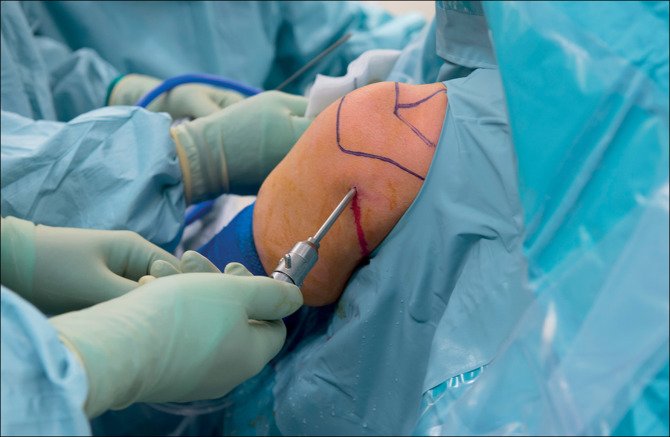
Procedure
The surgeon may inject fluid into the shoulder to expand the joint. This makes it easier to see all the structures in the shoulder through the arthroscope.
Next, the surgeon makes a small hole (about the size of a buttonhole) in your shoulder to insert the arthroscope. Fluid flows through the arthroscope to maintain visibility and control bleeding. Images from the arthroscope are projected onto a video screen to show the surgeon the inside of the shoulder and the injury.
Once the problem is clearly identified, the surgeon inserts additional small instruments through separate incisions to treat the problem. Specialized equipment is used for tasks such as shaving, cutting, grasping, suturing, and tying knots. Special devices are often used to secure the sutures to the bone.
Once the procedure is complete, the surgeon closes the incision with sutures or a steristrip (a small plaster) and covers it with a large, soft bandage.